Research

CNS Myelin
I'm a paragraph. Click here to add your own text and edit me. It’s easy. Just click “Edit Text” or double click me to add your own content and make changes to the font. Feel free to drag and drop me anywhere you like on your page.
I’m a great place for you to tell a story and let your users know a little more about you.
CNS Myelin
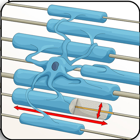
Nearly half of the nerves in the human brain and spinal cord are encased in a thick multilayered membrane - called a myelin sheath. Myelin sheaths, formed by glial cells known as oligodendrocytes, facilitate rapid signal propagation and maintain neuronal health. For decades the role of oligodendrocytes has been thought to simply maximize neuronal signaling speed. However, emerging evidence suggests that oligodendrocytes have a more interactive role in the nervous system, where myelin sheath size may tune neuronal signaling speeds to coordinate neural networks and, by being sensitive to neuronal activity, may be important for brain plasticity and learning. These roles are only beginning to be discovered.
Myelin sheaths are fundamental to a healthy nervous system. Multiple Sclerosis is the most prominent neurodegenerative disease resulting from myelin sheath loss. Many other neurodevelopmental diseases and disorders, psychiatric conditions, and neurodegenerative diseases throughout the lifespan are also associated with myelin alterations and loss.
There are many remaining questions about the impact of myelin sheath formation and maintenance to sculpting nervous system development, function, and contributions to disease. We aim to improve human health by understanding how myelin sheaths are formed and how their properties impact nervous system function.

Illustration use with permission from: © ScideLight
Cell Morphology


Form Meets Function
Is the morphology of an oligodendrocyte (i.e. the size of myelin sheaths) precisely regulated to provide an important function for the nervous system? For example, are the lengths of myelin sheaths key to signaling precision? While it has been known for decades that the size, particularly the length, of myelin sheaths determines the speed of signal propagation in axons, the overall contribution to neuronal circuitry is less well understood. Will achieving the appropriate size of myelin sheath be an important component for an effective strategy to restore function for those with neurodevelopmental and neurodegenerative disorders and diseases?
We are tackling these exciting questions.
At the heart of myelin sheath formation is the oligodendrocyte, the specialized myelin forming cells of the CNS. As they generate myelin sheaths, oligodendrocytes undergo an impressive change in cell size and shape. This creates a demand for coordinated intracellular transport and directional growth to enable an increase in membrane area up to several thousand-fold selectively at the tips of fine processes that wrap around neurons. By addressing the molecular signals and cellular machinery within the oligodendrocyte that coordinate the 3D membrane growth precisely and selectively around axons, we hope to uncover fundamental mechanisms that control cell shape and the mechanisms that control the formation, maintenance, and regeneration of myelin sheaths in the CNS.

We use a combination of bioengineered cell culture models, zebrafish, and mouse models to answer questions about
how myelin is formed during development and regeneration, and in turn, how the properties of myelin sheaths affect nervous system function.
We thank the generous support for our research from:


NIH NINDS
R01NS135206
